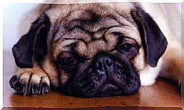
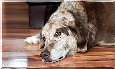
MRI In Pets
There are several methods that serve as tools for veterinarians to practice their profession. These techniques aim to facilitate the definitive diagnosis in the various clinical cases that appear for consultation. Undoubtedly, magnetic resonance imaging (MRI) is one of the most important.
Magnetic resonance imaging is a modern tool that became popular a few years ago, during which the excellent contributions it brings to the veterinary clinical field were observed. We invite you to continue this reading to discover how MRI performs in the diagnosis of pets.
What is magnetic resonance imaging (MRI)?
Magnetic resonance imaging is a diagnostic technique based on the use of magnetic fields and radiofrequency waves in the form of pulses, in which very clear images of the analyzed body parts are obtained. In addition, it allows you to observe the interior of tissues, which greatly facilitates the diagnosis of sick pets and humans.
Among the advantages of RM, the following stand out:
- Thanks to the use of tomographic slices, there is no overlapping of structures.
- Unlike X-rays and computed axial tomography, magnetic resonance does not use ionizing radiation, so it is considered an innocuous diagnostic alternative.
- Produces images in 2 and 3 dimensions in any plane, without the need to reposition or move the animal. Thus, it avoids causing any injury to the patient and obtains a better spatial resolution.
- It is not invasive or traumatic.
- Provides morphological and functional information.
- It allows a detailed observation of the composition of the lesions and provides an excellent contrast with the soft tissues.
- Does not cause pain in the patient.
MRI and veterinary medicine
Although MRI began in 1946, its popularity in medicine is relatively new. It is a technique that does not know races or species and has managed to promote an important development in the medical and surgical area of small living beings, from companion animals to wild fauna.
The main application areas in which magnetic resonance is used as a diagnostic method are as follows:
- Central and peripheral nervous system damage.
- Musculoskeletal and articular soft tissue injuries such as ligaments and tendons.
- Wounds in visceral areas – abdomen and chest.
- Presence of foreign bodies and vascular diseases.
How to prepare your pet for an MRI?
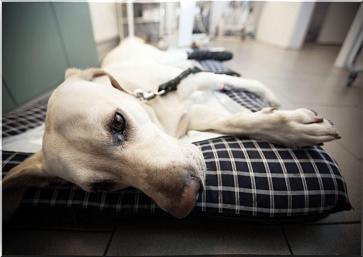
Magnetic resonance does not put the animal’s health at risk. However, it is important to consider some aspects before, during and after the exam.
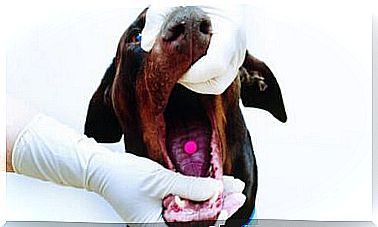
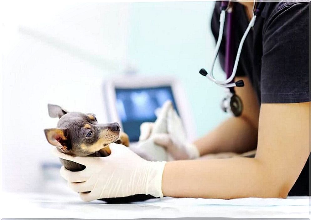
Its average duration is 30 to 90 minutes, during which the animal must remain immobile to obtain quality images. For this reason, it is necessary to administer anesthetics or, in some cases, tranquilizers.
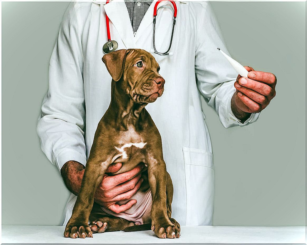
The decision will depend on the individual conditions of each patient. In order to make this choice, the animal will undergo a general physical and nervous evaluation, always with special emphasis on a routine pre-anesthetic analysis, in order to determine the ideal anesthetic protocol for him.
Once inside the room, the animal is placed on an examination table adapted for it. Then, a coil is placed around the area to be examined, which has the function of capturing radio waves. Finally, the animal is introduced into the magnetic tunnel.
In some patients, it is necessary to administer a paramagnetic contrast agent called gadolinium intravenously , which modifies the magnetic field and alters the signal intensity. This allows the veterinarian to better visualize the lesions and structures.
Can complications occur during an MRI?
As the animal will be subjected to a powerful magnetic field – which will cause an intense level of noise inside the room – it is advisable to put cotton in its ears. It is even possible to wear a special protective helmet.
On the other hand, if your pet has any metal parts on its body – such as screws, prostheses, staples, plates, microchips or pacemakers – it is important to let your veterinarian know.
All metals generate interactions with magnetic signals, which causes changes in the images obtained. In some cases, metals tend to become so hot from the action of magnetic force that they can cause secondary burns to the animal.
As with any other intervention that requires the use of anesthetics , there is a risk of complications during the process, such as heart failure or reaction to the medication administered. That’s why pre-anesthetic tests are so important to a patient’s health.
Although we are talking about a test that can be a little expensive, the main benefit that can be obtained from MRI is the reduction in the time between diagnosis and treatment of your pet, which means faster recovery and healing. .